
SEOUL, August 25 (AJP) - By easing off the chemical signals normally used to force stem cells into heart tissue, researchers at Sookmyung Women's University and Sungkyunkwan University have grown a model that lengthens and bends on its own, reproducing a stage of human heart development that has been almost impossible to observe.
Congenital heart disease is the most common birth defect in the world, occurring in about nine of every 1,000 live births since the mid-1990s, or roughly 1.35 million newborns a year, according to a systematic review published in the Journal of the American College of Cardiology. A later update covering 260 studies put Asia's rate highest among world regions, at about 9.3 per 1,000.
Most of those defects begin in the first weeks after conception, when the heart is a straight tube that has to stretch, curve and sort itself into an ordered sequence of future chambers. That window is effectively closed to observation. Human embryos at that stage are inaccessible, and animal hearts diverge enough from human ones to limit what mouse studies can settle.
Laboratory heart models have grown more sophisticated over the past five years, but most of them stay round. Researchers typically drive stem cells toward cardiac fate with strong external chemical signals, an approach that reliably produces beating heart tissue and just as reliably produces it as a sphere. Shape does not emerge, and neither does the ordering that depends on shape.
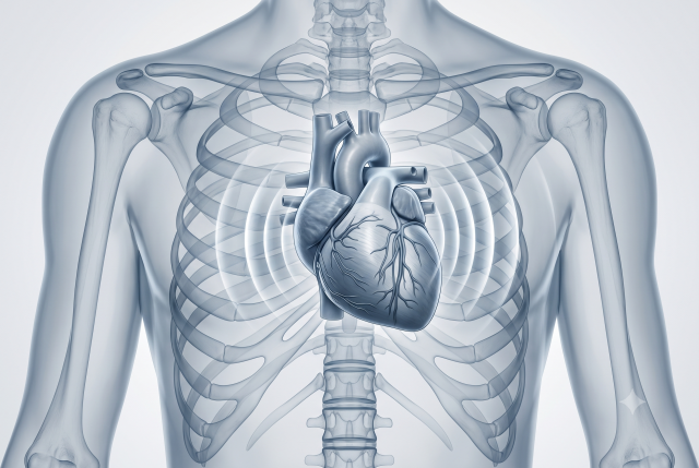
Grown from human induced pluripotent stem cells, adult cells reprogrammed back to an embryonic-like state, the resulting structures elongated and curved without being pushed into shape. The team named them elongating heart organoids.
Cells inside them settled along a venous-to-arterial axis in the order a developing heart follows, with sinus venosus-like cells at one end, then atrial cells, then ventricular cells. The sinus venosus is the inflow region of the embryonic heart and the site where the sinoatrial node, the heart's natural pacemaker, later forms.
The organoids also beat in sequence rather than all at once. Contractions started at the venous end and traveled toward the ventricular end, accompanied by a matching wave of calcium ions, the signal that triggers each contraction in heart muscle.
To work out what drove the lengthening, the team sequenced individual cells and ran trajectory analysis, a computational method that infers how cell populations relate to one another. That analysis pointed to dividing splanchnic mesoderm cells at the venous end, an early tissue layer that supplies cardiac cells in the embryo, feeding new cells into the structure as it grew. The inference comes from gene expression patterns. No one tracked individual cells making the journey.
The disease result came from deleting TBX5, a gene mutated in Holt-Oram syndrome and linked to septal and conduction defects in patients. Organoids missing it failed to elongate or bend, and contracted irregularly.
Bae claimed priority for the result. "This study is the first to reproduce the three-dimensional form and axis organization of the early heart outside the body by inducing the self-organizing capacity of stem cells," he said, adding that it should "raise the competitiveness of organoid-based drug development platforms and help establish the mechanisms behind congenital heart disease."
Other groups have built human heart models with patterned regions. Heart-forming organoids reported in Nature Biotechnology in 2021, multi-chamber cardioids in Cell in 2023, and a patterned primitive heart organoid in Nature Communications the same year each reproduce parts of early cardiac structure, and the Nature Communications model elongates and contorts as it grows. Reviews of the field describe most human systems as reaching heart tube formation without elongation. Whether the Sookmyung model is first in the narrower sense Bae describes cannot be checked against the university's summary alone.
Several things remain unshown. The organoids correspond to the earliest weeks of development and do not form chambers, septa, valves, coronary vessels, or the nerve tissue that regulates heartbeat. The university released no measurements, no culture duration, no count of organoids grown, and no success rate, leaving open how consistently the structures form. The TBX5 experiment establishes that removing the gene disrupts the model, not that the same sequence causes disease in patients. No drug has been tested in the system.
The work was carried out with researchers at AniMusCure Inc., a Suwon-based biotechnology company founded in April 2020. Bae and Kang are its co-chief executives. The company, whose existing pipeline covers sarcopenia and Duchenne muscular dystrophy rather than cardiac disease, plans to use the technology for congenital heart disease modeling, cardiac toxicity testing and screening cardiovascular drug candidates, the university said in announcing the work Aug. 20.
(Reference Information)
Journal/Source: Developmental Cell (IF 9.2, JCR top 3.95%)
Copyright ⓒ Aju Press All rights reserved.